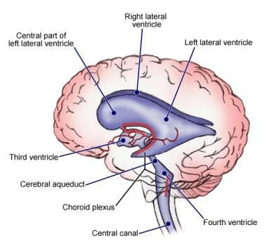


The metabolic waste of the brain is removed through an interstitial fluid that goes to the blood capillaries or into the ventricular system and can be recovered from the ventricular system by active reabsorption through the epithelium of the choroid back into the interstitial fluid and then into the blood capillaries or goes through the already mentioned absorption into the venous system ( 2).Īs the brain has no lymphatic capillaries, apart from the task of eliminating catabolic products, the cerebrospinal fluid also plays a role in the removal of macromolecules. Due to fibrosis of the interstitium in the choroid and oxidative damage to the epithelium, the transport of vitamins B and C is impaired. In the mature and old age, many of these nutrients do not reach the neurons in sufficient quantity. That is why liquor contains vitamins, peptides, nucleosides and growth factors. Unlike other organs that meet their metabolic needs through blood, the brain, due to the presence of the blood-brain barrier, does so through the cerebrospinal fluid. Passing through the ventricles, the CSF exits through the central orifice and lateral openings of the fourth ventricle, thus reaching the subarachnoid area or outer CSF ( 2). Interactions between the ANP, AVP, and FGF2 and their membrane receptors, located on the apical membrane of the cell, reduce the formation of CSF via neuroendocrine effects on the choroid.Īs the levels of ANP, AVP, and FGF2 are mainly under central control, such regulation is largely independent of the plasma composition or peripheral factors. Liquor formation, among other things, is influenced by the growth factors and neuropeptides by regulating the fluid exchange through the ependymus. Due to the energy-favorable gradients for Na+ (basally enters the cell) and K+, Cl-, HCO3- (apically exits the cell), a net flow of fluid and ions from the plasma, through the cell, into the CSF is generated. The second step takes place with the help of protein transporters located in the epithelium of the choroid. The first step takes place depending on the pressure difference between the plasma and the interstitial fluid of the choroid, so that, in cases of increased pressure of the cerebrospinal fluid, for example in hydrocephalus, the formation of the same is reduced. The formation can be divided into two phases: passive filtration of the fluid from the choroidal capillaries happens in the first stage, followed by active secretion via the choroid epithelium in the second ( 2). These spaces include ependymal cells lining the ventricles of the brain, but the arachnoid membrane, despite being able to secrete peptides and proteins, does not produce liquor. Most of it, about 100 mL, is formed in the choroidal mesh that is located in all four chambers and the rest, approximately 50 mL, is formed in other CNS spaces. It travels through the lateral ventricles (chambers) and through the interventricular openings to the third ventricle, then through the cerebral aqueduct to the fourth, from which it enters the subarachnoid space through lateral and median openings, and is then absorbed through the arachnoid fissures into the veins of the dura. Cerebrospinal Fluid and its FunctionĪccording to the classical theory, the cerebrospinal fluid is formed predominantly within the choroid segment, which is attached to the ventricular wall.

The lateral ventricles attach to the middle of the cerebral hemisphere and extend far into the forehead and the occipital region and into the temporal lobe ( 1).

Both of these ventricles are connected to the third ventricle by an intercellular (interventricular) opening. Inside both brain hemispheres there is a lateral ventricle, ventriculus lateralis. The lateral walls of the third cerebral ventricle form paired gray nucleus structures - the hill (thalamus) and the suburbs (hypothalamus). This “ water system” anteriorly expands to form the third ventricle, ventriculus tertius. The fourth ventricle continues into the narrow canal of the spinal cord.Īt the anterior end of the bridge, the fourth ventricle continues into a narrow tube that runs into the midbrain and is also called the water system of the midbrain ( 1). The elongated brain and bridge form a rhombic pit, forming the bottom of the fourth brain ventricle, ventriculus quartus, and its roof, like a vault, forms the cerebellum.


 0 kommentar(er)
